Quick answer: NICE uses the Canadian C-spine rule to decide who needs cervical spine imaging after trauma. Any high-risk factor (age 65 or over, a dangerous mechanism, or paraesthesia in the limbs) means immobilise and image. If there is a low-risk factor, ask the patient to actively rotate the neck 45 degrees left and right: if they can, no imaging is needed. In adults the imaging of choice is CT. In children it is MRI if there is a strong suspicion of injury.
Cervical spine assessment appears regularly in the MRCEM SBA, usually as a trauma scenario asking whether the patient needs imaging, which scan to choose, or whether the collar can come off. This guide follows NICE guideline NG41, Spinal injury: assessment and initial management.
First steps in any trauma patient
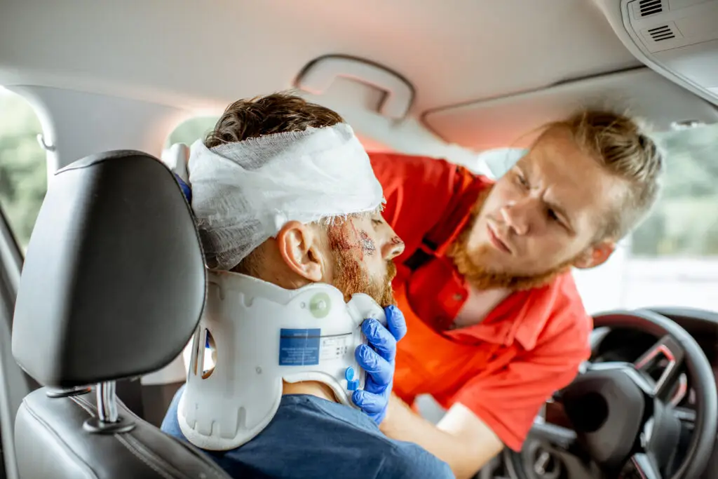
- Protect the cervical spine with manual in-line stabilisation while you assess, particularly during airway management.
- Assess for spinal injury as part of the primary survey, and check whether the patient has any factor that makes assessment unreliable, such as reduced consciousness, intoxication or a distracting injury.
- If the assessment cannot be completed, maintain full in-line spinal immobilisation.
The Canadian C-spine rule
Step 1: is there a high-risk factor?
The patient is at high risk if any one of these is present:
- Age 65 years or older.
- Dangerous mechanism of injury: a fall from a height of more than 1 metre or 5 steps, an axial load to the head (for example diving), a high-speed motor vehicle collision, a rollover, ejection from a vehicle, an accident involving motorised recreational vehicles, a bicycle collision or a horse riding accident.
- Paraesthesia in the upper or lower limbs.
Any high-risk factor means the patient needs full immobilisation and imaging.
Step 2: is there a low-risk factor?
If there are no high-risk factors, NICE classes the patient as low risk if they have one of the following:
- Involved in a minor rear-end motor vehicle collision.
- Comfortable in a sitting position.
- Ambulatory at any time since the injury.
- No midline cervical spine tenderness.
- Delayed onset of neck pain.
A low-risk factor means it is safe to go on and assess neck movement.
Step 3: can the patient rotate the neck?
Ask the patient to actively rotate the neck 45 degrees to the left and to the right.
- Able to rotate and pain free: NICE classes the patient as no risk. No imaging or immobilisation is needed.
- Unable to rotate: the patient remains at low risk. Maintain immobilisation and image.
| Finding | Action |
|---|---|
| Any high-risk factor | Immobilise and image |
| Low-risk factor present, but cannot rotate 45 degrees each way | Immobilise and image |
| Low-risk factor present and can rotate 45 degrees each way, pain free | No imaging needed |
Which imaging?
Adults (16 and over)
- CT is the first-line investigation when imaging is indicated by the Canadian C-spine rule.
- If there is a neurological abnormality that could be due to spinal cord injury, perform MRI after CT, whether or not the CT shows an abnormality.
Children (under 16)
- Perform MRI if there is a strong suspicion of cervical spinal cord injury (from the Canadian C-spine rule and clinical assessment) or of spinal column injury (from clinical assessment or abnormal neurological signs or symptoms).
- Consider plain X-rays in children who do not meet the criteria for MRI but in whom clinical suspicion remains after repeated assessment.
Children can have a spinal cord injury with normal X-rays and CT, known as SCIWORA (spinal cord injury without radiological abnormality). This is one reason MRI is preferred.
Do not forget the thoracic and lumbar spine
Suspect a thoracic or lumbosacral injury if any of the following is present:
- Age 65 or older and reported pain in the thoracic or lumbosacral spine.
- Dangerous mechanism: a fall from a height of more than 3 metres, or an axial load to the head or base of the spine.
- Pre-existing spinal pathology, or known or at risk of osteoporosis.
- Suspected spinal fracture in another region of the spine.
- Abnormal neurological symptoms or signs.
- On examination: new deformity, bony midline tenderness, or midline or spinal pain on coughing.
A fracture at one spinal level increases the chance of a second fracture elsewhere, so image the whole spine when one injury is found.
Spinal cord injury: features to recognise
- Neurogenic shock: hypotension with bradycardia and warm peripheries, caused by loss of sympathetic tone in injuries at or above T6. Exclude haemorrhage first, as it is the commonest cause of shock in trauma.
- Spinal shock: temporary flaccid paralysis and loss of reflexes below the level of injury. It is a neurological phenomenon, not a circulatory one.
- High cervical injury (C3–C5) can cause respiratory failure through loss of diaphragmatic function.
Patients with a suspected spinal cord injury should be discussed early with the spinal cord injury centre.
Common exam traps
- Testing neck movement in a high-risk patient: anyone aged 65 or over, or with a dangerous mechanism or paraesthesia, goes straight to imaging.
- Choosing X-rays in an adult: CT is first line in adults.
- Choosing CT in a child: MRI is preferred when suspicion is strong.
- Stopping at a normal CT when there are neurological signs: MRI is still needed.
- Confusing the heights: more than 1 metre or 5 steps for the cervical spine, more than 3 metres for the thoracic and lumbar spine.
- Blaming hypotension on neurogenic shock: look for bleeding first.
Summary
- NICE uses the Canadian C-spine rule: high-risk factors, low-risk factors, then active rotation to 45 degrees.
- High-risk factors: age 65 or over, dangerous mechanism, limb paraesthesia.
- Adults: CT, with MRI as well if there are neurological signs.
- Children: MRI if suspicion is strong, and consider X-rays if suspicion persists but MRI criteria are not met.
- Check the thoracic and lumbar spine too.
Frequently asked questions
What are the high-risk factors in the Canadian C-spine rule?
Age 65 or older, a dangerous mechanism of injury, and paraesthesia in the upper or lower limbs.
When can the cervical spine be cleared without imaging?
When there are no high-risk factors, at least one low-risk factor is present, and the patient can actively rotate the neck 45 degrees left and right without pain.
What is the first-line cervical spine imaging in adults?
CT. MRI is added if there is a neurological abnormality that could be due to spinal cord injury.
What imaging is used for suspected cervical spine injury in children?
MRI when there is a strong suspicion of injury. Plain X-rays are considered when the MRI criteria are not met but clinical suspicion remains.
Next steps
Try our 20 free MRCEM SBA sample questions to test yourself. For more, explore our Trauma questions and the Trauma course, or see the full question bank and mock tests in our membership plans.
More MRCEM clinical topics: Head injury CT criteria, Sepsis in the ED, Suspected pulmonary embolism, Chest pain and ACS.
This article is for exam preparation. In clinical practice, always follow current NICE guidance and your local trauma protocol.